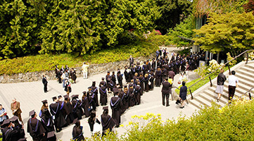
Congratulations Biochem graduates!! May Graduation, we have 2 separate days to celebrate your achievements. UBC will be conferring:
MSc and PhD degrees on Wednesday, May 23rd at 8:30 am
BSc degrees will be held on Thursday, May 31st at 11:00 am
Congratulations Biochem graduates!! May Graduation, we have 2 separate days to celebrate your achievements. UBC will be conferring:
MSc and PhD degrees on Wednesday, May 23rd at 8:30 am
BSc degrees will be held on Thursday, May 31st at 11:00 am
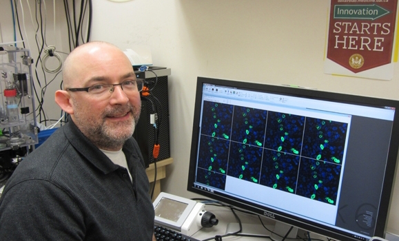
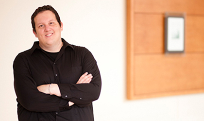
 Canadian Society for Molecular Biosciences, (CMSB) awards Filip van Petegem CSMB New Investigator Award. Filip’s cutting-edge structural studies have provided fundamental insights into our understanding of integral membrane proteins, including ryanodine receptors and voltage-gated sodium channels. His structure function studies comparing wildtype and disease mutant forms of membrane proteins have provided valuable insight into a large number of genetic diseases as well as fundamental, mechanistic understandings of ion channel functions.
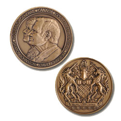
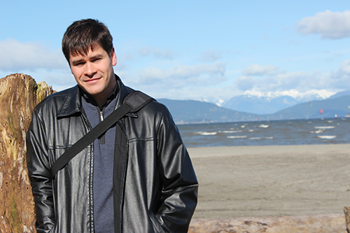 Dustin King, PhD, trainee from Natalie Strynadka’s Lab has been selected to receive a Governor General’s Gold Medal. Only one Gold Medal is awarded to the student who has achieved the most outstanding academic record as a doctoral student. Dustin is one of 300 PhD’s graduating from UBC this year. Medals are presented on behalf of the Governor General of Canada by participating educational institutions, along with a personalized certificate signed by the Governor General. There is no monetary award associated with the medals.
 “CRISPR advances in genome editing,” by Graham Dellaire, Cameron Research Scientist in Cancer Biology, Associate Professor, Department of Pathology, Dalhousie University.
Monday, April 25, 2016, LSC #3 at 3:00 pm, 2350 Health Science Mall
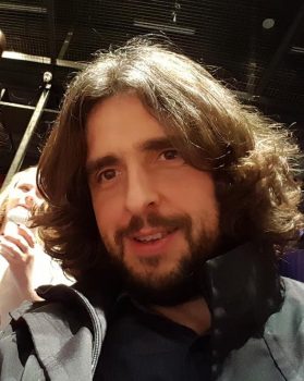
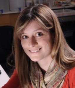
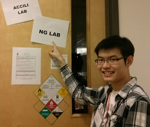 Leo Ng, a PhD student in Cell and Development Biology and a trainee in Dr. Accili’s Lab, is awarded a Killam Graduate Teaching Assistant Award for TA’ing Biochemistry undergraduate courses. Leo has been a TA for us since 2013 Winter Session. Over the years, the comments in his TA evaluations are always positive and remarks on his valuable contribution to our student’s education. Congratulations Leo!
This award is by nomination across UBC campus. In recognition of the valuable role that teaching assistants play in our undergraduate programs, UBC annually awards teaching prizes to sixteen UBC Teaching Assistants. The prize includes a certificate and $1,000. Successful candidates will have met criteria that result in a high level of respect from undergraduate students and academic or course supervisors.
The awards, given annually since 1996/1997, send the important message to students (both undergraduate and graduate) that UBC values teaching and seeks to recognize teaching excellence at our university.
https://academic.ubc.ca/awards-funding/award-winners/teaching-service-award-winners
 “Network analysis in the prediction of proteolytic pathways,” by Nikolaus Fortelny, PhD candidate, Overall and Pavlidis labs, UBC.
Monday, April 18, 2016, LSC #3 at 3: 00 pm, 2350 Health Science Mall
 “Molecular Promiscuity and Specificity: The Far-from-perfect Enzyme and the Struggle for Survival”, by Mikael Elias, Assistant Professor, Department of Biochemistry, Molecular Biology, and Biophysics, University of Minnesota.
Monday, April 11, 2016, LSC 3 at 3:00 pm, 2350 Health Sciences Mall.
“Generation of Truncated Proteoforms in Proteolytic Networks: Modelling and Prediction in the Protease Web”, by Nikolaus Fortelny. Wednesday, April 20, 2016 at 4:00 PM in Room 203 of the Graduate Student Centre, 6371 Crescent Road
 “An Evolutionary Module in Central Metabolism”, Kimberly Reynolds, Assistant Professor, Green Center for Sytems Biology, Department of Biophysics, University of Texas Southwestern.
Monday, March 7, 2016, LSC 3 at 3:00 pm, 2350 Health Sciences Mall
 Linking Metabolic Activities to Chromatin Functions in Epigenetic Processes, presented by Lorraine Pillus, Professor and Chair, Division of Biological Sciences, University of California, San Diego.
Monday, February 22, 2016 LSC 3 at 3:00 pm, 2350 Health Sciences Mall