Posted on January 20, 2022
Title: Structural basis for the DASS/SLC13 family’s carboxylate transport, chemical inhibition, and pathogenesis
Dr. David Sauer, P.I of Membrane Protein Structural and Chemical Biology, Centre for Medicines Discovery, University of Oxford.
Abstract: Citrate, α-ketoglutarate, and succinate are TCA cycle intermediates that also play essential roles in metabolic signalling and cellular regulation. These molecules are imported across the plasma membrane by the divalent anion sodium symporter (DASS) protein family, encoded by the SLC13 genes in humans. Underlining their biomedical importance, mutations in NaDC3 (SLC13A3) and NaCT (SLC13A5) result in neurological disorders. We determined several structures of two bacterial DASS proteins, including the previously unseen outward-facing state, to provide biophysical descriptions of the family’s elevator transport mechanism and strict substrate coupling. Building from this, we determined structures of the human citrate transporter NaCT in complexes with citrate or a small-molecule inhibitor. These revealed how the inhibitor arrests the transport cycle of NaCT, and explains compound selectivity. Together, the bacterial and NaCT structures provide a framework for understanding how various mutations abolish the transport activity and thereby cause SLC13A5-Epilepsy.
Monday, January 31, 2022 at 2:30 pm via Zoom
Hosted by: Dr. Seth Parker
Read More | No Comments
Posted on January 6, 2022
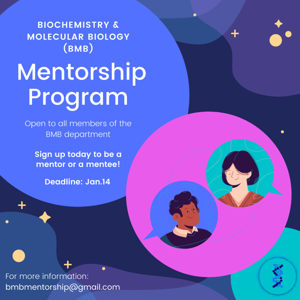
The Biochemistry & Molecular Biology (BMB) department is proud to introduce a new mentorship program that will be piloted this upcoming term. The program will match mentors and mentees within BMB to build meaningful connections with a professional, career, and scientific focus.
This program is open to all members of the BMB department (Undergrads, Grad students, post-Docs, PI’s) and will be running from Jan-Apr 2022. This initiative aims to foster a sense of community within the BMB program with an emphasis on Equity, Diversity, and Inclusion.
Read More | No Comments
Posted on December 29, 2021
Read More | No Comments
Posted on December 21, 2021
Read More | No Comments
Posted on December 15, 2021
Read More | No Comments
Posted on December 6, 2021
Read More | No Comments
Posted on December 1, 2021
Title: “Structural characterization of the Type 3 secretion system needle complex by single particle cryogenic electron microscopy”
Abstract: “The bacterial type III secretion system, or injectisome, is a syringe-shaped nanomachine essential for the virulence of many pathogenic Gram-negative bacteria. A major functional subcomplex of the injectisome, the needle complex, is a 3.5MDa complex formed by more than ten unique proteins. The needle complex forms a continuous channel spanning both the inner and outer membranes of Gram-negative pathogens, created by three highly oligomerized inner and outer membrane hollow rings and a polymerized helical needle filament. The effector proteins secreted through this channel, which vary amongst different bacterial species, are essential for subsequent pathogenicity. Thus, structural studies of this complex can provide important atomic level information for understanding complex assembly and function of the injectisome as well as potentially development of new antivirulence drugs or vaccines to combat infections in susceptible human, animal and plant hosts pathogens by developing new drugs or improve the existed ones.
The first high-resolution needle complex structures determined by cryogenic electron microscopy (cryo-EM) here shows the atomic details of the inner and outer membrane protein complex and the needle filaments. The outer membrane component of the needle complex belongs to the secretin family, a giant necessarily gated pore common and essential to other bacterial secretion systems but which had remained largely uncharacterized at the atomic level until recent work including major contributions as outlined in this thesis. Notably, the structures of the “open” conformation of the type III secretion system secretin and the needle filament “substrate” which passes through its inner channel revealed the gating mechanism for the first time in the secretin family. Further, the structures of the dual nested rings that form the major inner membrane structural component of the needle complex shows remarkable similarity regardless of the assembly stages, inferring a highly stable foundation for the other components of the system to pack and function within. The snapshots of multiple needle complexes at different assembly stages revealed multiple new structures, the dynamics of the assembly, and showed the sequence of the assembly process. The structural information also answered several long-standing additional questions, such as the mystery of the apparent symmetry mismatch between the inner and outer membrane complex of prior structures, and the fold, span and functional role of the historically named “inner rod” protein which this thesis works shows is not a rod at all but an adaptor to set the needle helicity and anchor it to the stable inner membrane platform.”
Monday, December 13, 2021 at 2:30 pm at LSC #3 and or join by Zoom.
Hosted by: Dr. Natalie Strynadka
Read More | No Comments